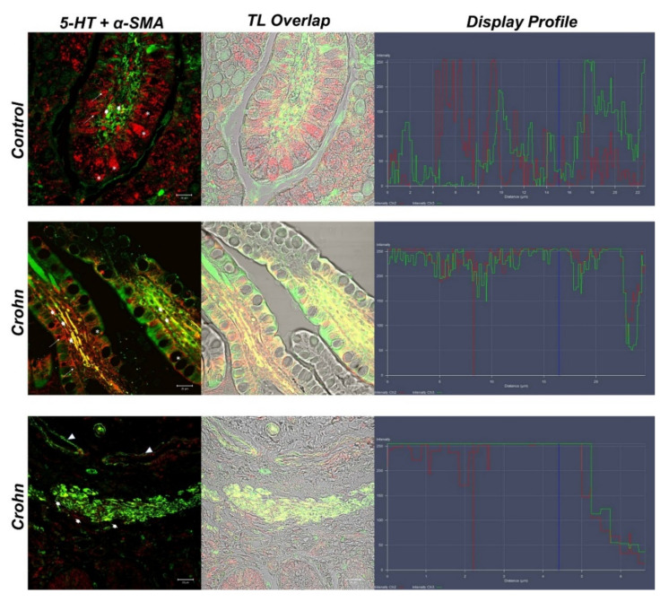
Figure 3.
Human intestine, 5-HT/α-SMA colocalization, 20×, scale bar 20 nm. Merge photomicrographs indicate the double localization of two antibodies in myofibroblasts, in which it is evident that the overlap (yellow fluorescence) concerns the CD samples rather than in the healthy intestine. In the control section 5-HT highlighted the goblet cells cytoplasm (*). A cluster of subepithelial myofibroblasts in the lamina propria colocalized with 5-HT and α-SMA (large arrows), blood vessels (arrowheads). The graph represents the “display profile” function of the laser scanning microscope to show the intensity profile of detected antibodies.

