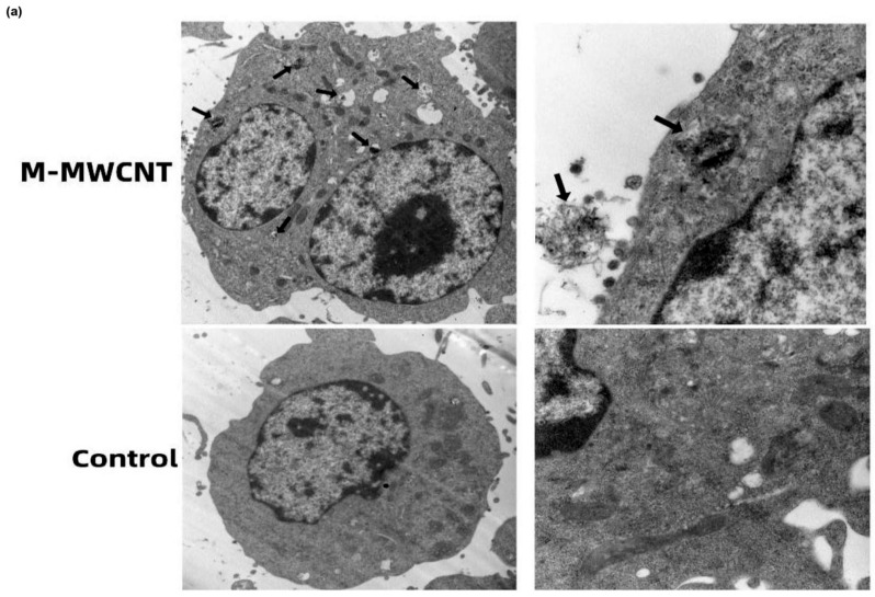
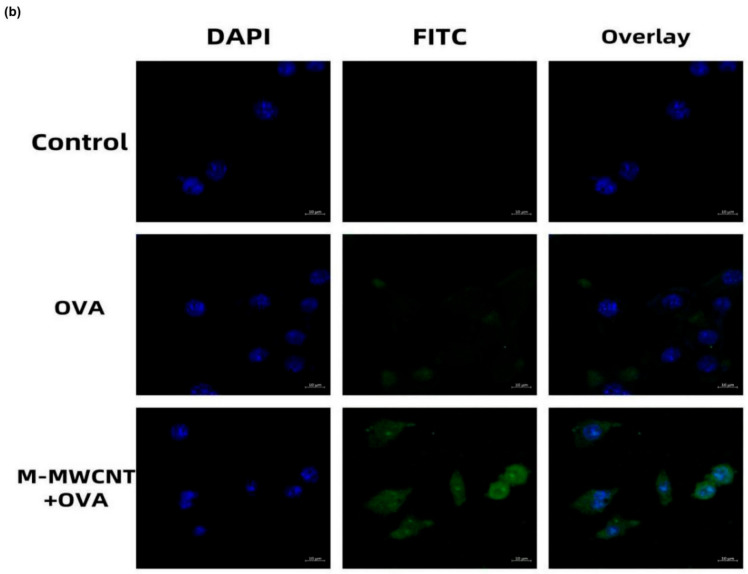
Figure 6.
CLSM and TEM image of M-MWCNT uptake by RAW267.4. (a) TEM micrographs of M-MWCNT (indicated by arrow) internalized by macrophages. M-MWCNT was co-cultured with macrophages for 12 h and then the collected cells were immediately fixed with 2.5% glutaraldehyde. The fixed cells were sectioned and observed by TEM. (b) CLSM image of FITC-OVA uptake by RAW267.4. Macrophages were cultured with M-MWCNT+FITC-OVA or FITC-OVA for 24 h. The slides were fixed and stained with DAPI as indicated by blue fluorescence, while green fluorescence indicates FITC-OVA. The cells were observed and photographed with a laser confocal scanning microscope.


