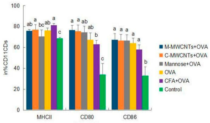
Figure 7.
Effects of M-MWCNT on the maturation of DCs in ICR mice. The splenocytes suspension were prepared from the spleens of ICR mice 48 h after the first immunization. Point diagram analysis of dendritic cell double-stained with CD11c+-MHC-II+, CD11c+-CD86+, and CD11c+-CD80+. The percentage of CD80+, MHC II+, and CD86+ cells against total DCs was determined by using flow cytometry. Distribution map of CD11c+-MHCII+, CD11c+-CD86+, and CD11c+-CD80+ co-expressing cells in Supplementary Materials. The data are presented as the mean ± SD (n = 3) analyzed by one-way ANOVA and then Duncan’s multiple-range tests. Different letters (a, b, c, and d) above each group of bars indicate statistically significant differences (p < 0.05).

