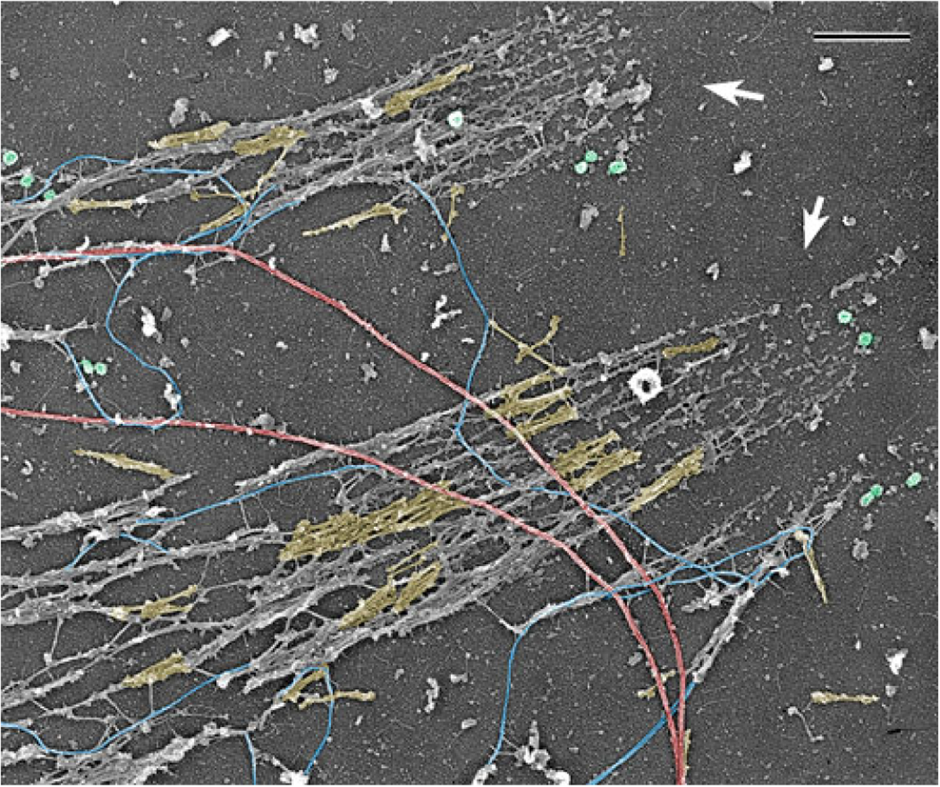
Fig. 3.

PREM of the cytoskeleton of a cultured PtK1 potoroo epithelial cell after depletion of actin filaments by gelsolin treatment. Image shows a peripheral cell region containing two large focal adhesions (arrows). Abundant myosin II filaments, a subset of which is highlighted in yellow, accumulate along the actin-depleted stress fibers extending away from focal adhesions, while some individual myosin II filaments can be seen elsewhere in the cytoplasm. Exposed microtubules (red), intermediate filaments (blue), and clathrin-coated vesicles (green) are also visible. Scale bar: 500 nm
