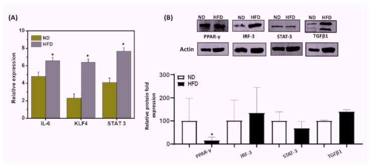
Figure 6.
Effects of HFD or ND feeding on mRNA expression and protein levels in mouse AT. Mice were fed HFD or ND for 12 weeks before sacrifice, and AT was collected from each group. (A) RT-qPCR analysis of IL-6, KLF-4, and STAT3 transcripts in AT was obtained from mice fed with ND or HFD. Total RNA was isolated from AT from each group of mice, pooled, quantitated, reverse-transcribed into cDNA, and analyzed by qPCR with primers specific for IL-6, KLF-4, and STAT3. HFD mice expressed higher levels of IL-6, KLF-4, and STAT3 transcripts than did mice fed ND. (B) Immunoblot analysis of PPAR-γ, TGFβ1, IRF3, and STAT3 protein expression levels in mice fed for 12 weeks with ND. AT from mice fed HFD or ND was lysed to obtain whole cell lysates and relative levels of TGFβ1, IRF3, PPAR-γ, and STAT3; proteins were determined by immunoblot analysis using specific monoclonal antibodies. HFD mice exhibited higher levels of protein expression of TGFβ1 and IRF3 than did mice fed ND. In contrast, protein expression levels of PPAR-γ and STAT3 were decreased in HFD-mice relative to the ND control. Vertical bars represent mean SEM, and significant differences between the groups are shown with (*) (p < 0.01) based on one-way ANOVA.

