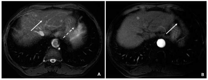
Figure 4.
Initial MRI with normal AFP and elevated %L3 (July 2008). (A) The axial T2-weighted gradient echo image shows a small hyperintense lesion (arrow) in segment 4A. A second hyperintensity (dashed arrow) corresponds to a hemangioma. (B) The corresponding axial T1-weighted fat-suppressed arterial-phase postcontrast image shows hyperenhancement (arrow) in the lesion subsequently found to be a small HCC, although currently without associated features to clinch the diagnosis. Without washout, capsule appearance, or other typical HCC findings, the lesion is indeterminate.

