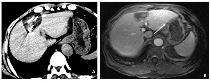
Figure 6.
Cryoablation of the segment 4A HCC (April and May 2009). (A) The CT image through the region of the segment 4A HCC shows the cryoprobes and formation of the surrounding ice ball that accumulates as freezing cycles are applied to ablate the lesion. (B) A delayed postcontrast T1-weighted fat-suppressed image 1 month later shows little to no apparent enhancement in the ablated lesion (arrow).

