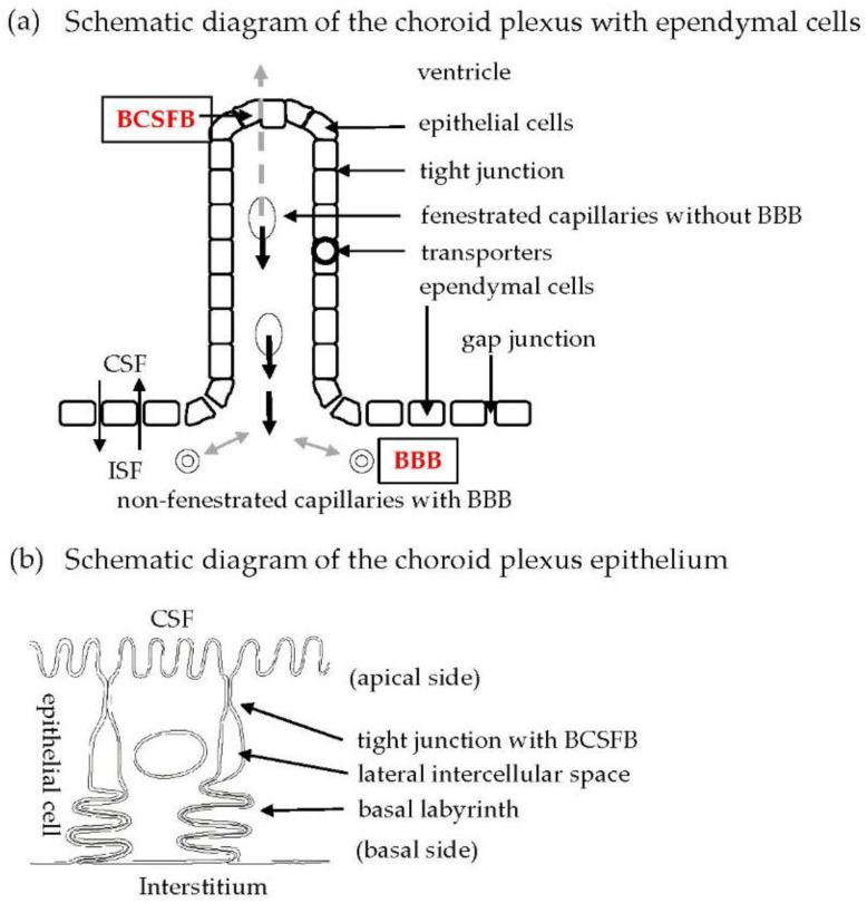
Figure 1.
(a) Schematic representation of BCSFB in CPE cells. Fenestrated capillaries are located in the stroma of CP. CPE cells facing the ventricle are bound by tight junctions. Ependymal cells mainly bound by gap junctions are located between the ventricle and brain parenchyma. Transporters are localized in the cytoplasmic membrane of CPE cells. Non-fenestrated capillaries with tight junctions between endothelial cells are situated in the brain parenchyma and have a tight barrier function, referred to as the blood–brain barrier (BBB). (b) Schematic diagram showing localization of the tight junction, lateral intercellular space, and basal labyrinth on the lateral side of CPE cells.

