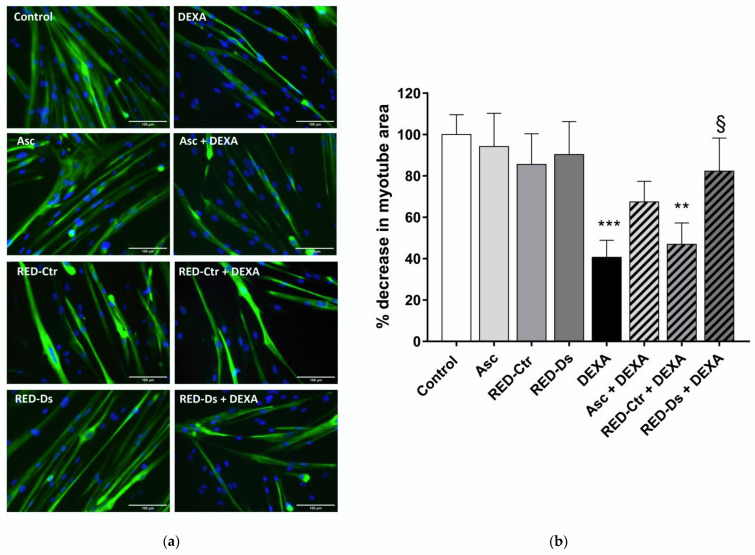
Figure 6.
Tomato peel extract effect on myotube atrophy. (a) Representative images of differentiated myotubes (MYH2-positive cells (green), with Hoechst-labeled nuclei in blue) after 48 h of treatment with 50 µM DEXA or 5 µg/mL TPC tomato peel extracts (RED) of plants grown in normal (Ctr) or drought stress (Ds) conditions. Ascorbic acid (Asc) was used as positive control. All images at ×20 magnification; scale bar = 100 microns. (b) Plot shows the percent decrease, from untreated cultures, in myotube area at each treatment condition. Data represent means ± SEM, n = 10. ** p < 0.005 and *** p < 0.001 vs. untreated cell (control); § p < 0.05 vs. DEXA using one-way ANOVA.

