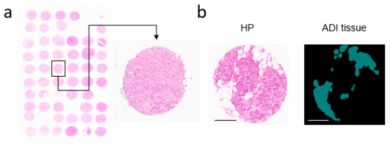
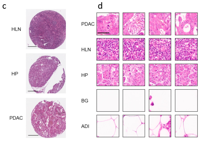
Figure 1.
Data Pre-processing Pipeline for H&E-stained Tissue Micro Arrays provide datasets for Deep Transfer Learning. (a) Patients’ data were extracted from tissue micro arrays (TMAs) and annotated. (b) A representative HP Spot with adipose tissue and a segmentation of the adipose tissue are shown (scale bar = 300 µm). (c) Spots from healthy lymph node (HLN), healthy pancreas (HP) and pancreatic ductal adenocarcinoma (PDAC) (scale bar = 300 µm). (d) Whole images were cut into square patches with 224 × 224 pixel sizes (scale bar = 60 µm). PDAC, HLN, HP, Background (BG), and Adipose Tissue (ADI) sample image tiles are shown.


