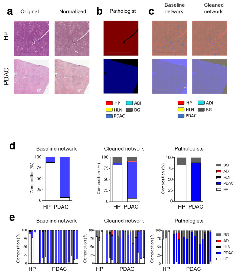
Figure 4.
Convolutional Neural Network can classify healthy pancreas tissue and pancreatic ductal adenocarcinoma. (a) Sections from independent H&E-stained whole images from healthy pancreases (HP) and pancreatic ductal adenocarcinoma (PDAC) are shown (scale bar = 2 mm). (b) Expert label (ground truth), as determined by a pathologist and (c) classified with the baseline and cleaned network, are shown. HLN (yellow), HP (red), PDAC (blue), background (BG, grey) and adipose tissue (ADI, cyan) are indicated by the pathologist (b) or the CNNs (c). The pooled (d) and individual classification (e), as determined using a baseline and cleaned network, as well as by a pathologist, of whole-image slides from healthy pancreases (HP) (n = 3) and pancreatic ductal adenocarcinoma (PDAC) (n = 15) are shown.

