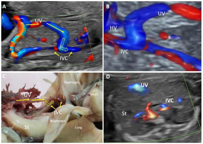
Figure 7.
Agenesis of the ductus venosus with a wide extrahepatic shunt to the inferior vena cava at 20 weeks of gestation (case 11). (A): Longitudinal plane of the fetal abdomen, color Doppler evaluation, showing the absence of ductus venosus and a wide umbilical shunt directed to the inferior vena cava. (B): 3D evaluation shows abnormal umbilical vein drainage into the inferior vena cava. (C): Pathological examination showing the drainage of the umbilical vein into the inferior vena cava. (D): Transverse view at the level of insertion of the umbilical cord showing the absence of the portal venous system and persistent small stomach (white arrow) UV—umbilical vein. IVC—inferior vena cava. HV—hepatic vein.

