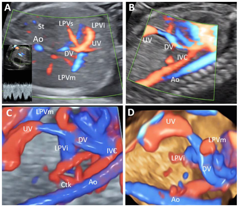
Figure 9.
Partial portal venous system agenesis (PPVSA) (Case 13–20 weeks of gestation). (A): Transverse view of the fetal abdomen, showing the confluence of the umbilical vein (UV) with left portal vein (LPV) branches, but the absence of a normal portal sinus and right portal vein. (B): Sagittal view showing the presence of ductus venosus. (C,D): Sagittal view showing in 3D the presence of ductus venosus and left portal vein branches. Ao—aorta; IVC, inferior vena cava; LPVi—left portal vein inferior branch; LPVm—left portal vein medial branch; LPVs—left portal vein superior branch; UV—umbilical vein; DV—ductus venosus; Ctk—celiac trunk; St—stomach.

