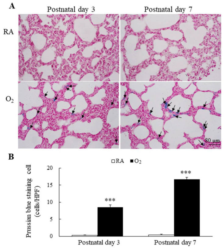
Figure 3.
(A) Representative photomicrographs of Prussian blue staining and (B) the positive cells per high-power field in lung tissue. Prussian blue staining was mainly localized in type II alveolar cells and alveolar macrophages (black arrow). The mice reared in hyperoxia exhibited a significantly higher Prussian blue positive staining cells per high-power field than the mice reared in RA. n = 6 mice at each postnatal day. *** p < 0.001.

