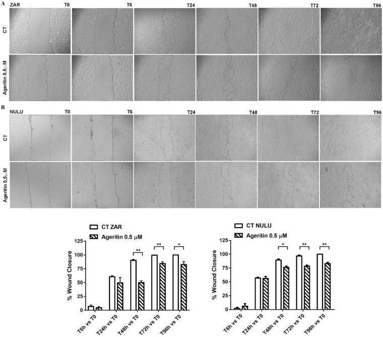
Figure 3.
Wound healing assay on primary glioblastoma cell lines NULU and ZAR under treatment with Ageritin (0.5 μM). (A,B) Representative images of ZAR and NULU cell wound healing at 0, 6, 24, 48, 72 and 96 h, under Evos FL microscope (4× magnification). Graphs reported the quantification of the wound healing assays as percentage of wound closure. Data are reported as mean ± SEM of 3 individual determinations. Unpaired t-test, p-value < 0.05. According to GraphPad Prism 7, * p-value 0.01 to 0.05 (significant), ** p-value 0.001 to 0.01 (very significant).

