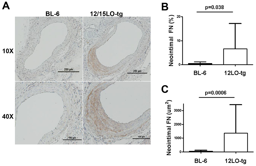
Figure 6. 12/15LO-tg mice have greater neointimal fibronectin (FN) compared with BL-6 in 28-day post-injury neointima.

To examine whether enhanced fibronectin deposition contributed to the increased neointimal formation in the 12/15LO-tg mice, we stained the 28D post-injury LCCA sections for fibronectin. 6A: top row displays 10X magnification of the representative sections stained for fibronectin in BL-6 and 12/15LO-tg mice. The bottom row displays the same slides at 40X magnification. 6B and C: show that compared with the BL-6, neointima in the 12/15LO-tg mice had significantly more fibronectin determined by the absolute fibronectin-stained area and by the percent of the fibronectin-stained area to the neointimal lesion. Data is represented as mean ± SD of the mean of two LCCA sections obtained from equally distributed intervals from carotid bifurcation of eight animals in each group.
