Figure 2.
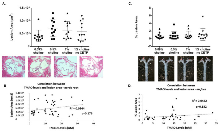
Effect of TMAO production on aortic lesions in vivo. Apoe−/− mice expressing CETP or controls (no CETP) were fed chow with supplementation of low, moderate and high choline for 16 weeks. Following 16 weeks of treatment, animals were sacrificed for aortic lesion assessments. (A) Sections (~10 μm) of the aortic root were cut, followed by hematoxylin–eosin staining (images below graph) and subsequent Nikon imaging. The total lesion area (μm2) across the aortic root for each treatment group was then plotted as the mean ± SEM from 11–12 animals per group. (B) Pearson correlation analysis was performed between aortic root lesion area and 16-week plasma TMAO concentrations for all groups (n = 43 mice). (C) Thoracic aortas were isolated, formalin-fixed and stained with Sudan IV for en face analysis (images under graph) using a Nikon computerized imaging system. The % lesion area in thoracic aortas for each treatment group was then expressed as the mean ± SEM from 11–12 animals/group. (D) Pearson correlation analysis was performed between % lesion area and 16-week plasma TMAO concentrations for all groups (n = 43 mice). One-factor ANOVA test was used to analyse normally distributed data, and a Kruskal–Wallis test was conducted for non-normally distributed data. Post hoc Tukey’s honestly significant difference test (HSD) pairwise comparisons were conducted to identify statistical differences between the treatment groups when a significant main effect was found. TMAO: trimethylamine N-oxide; CETP: cholesterol ester transfer protein.

