Figure 5.
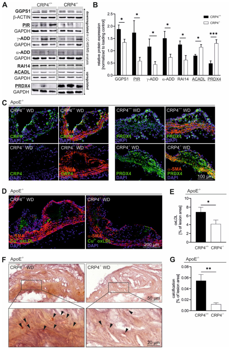
Vascular CRP4 regulates the expression of several proteins playing key roles in “oxidation reduction” and in atherosclerotic plaque formation. (A,B) Significantly regulated proteins identified in LC-MS/MS proteomic analysis (Table 1) of CRP4 WT and KO synthetic VSMCs were validated applying immunoblot technique. GetGo analysis indicated an enrichment of proteins involved in “oxidation reduction” (GO: 0055114) (underlined). Geranylgeranyl diphosphate synthase 1 (GGPS1), retinoic acid induced protein (RAI) 14, alpha- and gamma-Adducin (α- and γ-ADD) and Pirin (PIR) were significantly depleted in protein lysates obtained from P10-15 VSMCs lacking CRP4, whereas peroxiredoxin (PRDX) 4 and acyl-CoA dehydrogenase long chain (ACADL) were confirmed as significantly upregulated proteins in CRP4 KO, GAPDH and β-actin were co-detected to demonstrate equal loading of the gels; n = 7–11 per genotype where n represents a pool of four aorta, * p < 0.05, *** p < 0.001 and two-tailed Student’s t-test. (C) Immunofluorescence-based co-detection of CRP4 (green) and PRDX4 (green) as well as α-SMA (red) in frozen aortic tissue sections. CRP4 (left panel) was present in media and in intra-plaque cells, and it was detected in the fibrous cap of CRP4+/+ but not CRP4−/−. PRDX4 (right panels) was expressed in medial and plaque cells, and it was significantly upregulated in dKO atherosclerotic plaques and the underlying media. Intra-plaque PRDX4 did not overlap with α-SMA suggesting that these cells derived from a non-VSMC source or that these cells resemble VSM-like cells that lost α-SMA during dedifferentiation; n = 4 to 5 for the CRP4 and PRDX4 staining per genotype. (D,E) OxLDL was visualized using a Cu2+ oxLDL antibody (green). A lack of CRP4 was associated with a significant lower oxLDL content in atherosclerotic plaques. Co-staining with α-SMA (red) was performed to better clarify the localization of oxLDL within the different plaque structures; n = 6–7 per genotype, * p < 0.05 and two-tailed Student’s t-test. (F,G) Alizarin staining of aortic cryosections identified a potentially protective plaque-calcification defect in dKO compared to ApoE−/−/CRP4+/+ lesions; n = 7 per genotype, ** p < 0.01 and two-tailed Student’s t-test. Data shown in (B,E,G) are expressed as the mean ± SEM.

