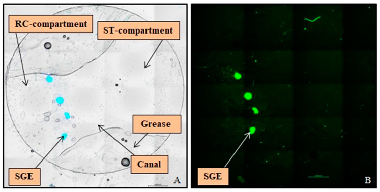
Figure 4.
Overview pictures of Spiral Ganglion Explants (SGEs) on the Ø9 mm Glass Coverslip after Immunocytochemistry. (A) In the merged bright field image four SGEs stained against DAPI (blue) are visible, cultured on the bottom of the Rosenthal’s canal-compartment in front of the canal. The used grease for sealing the Neurite Outgrowth Chamber left an imprint on the glass coverslip. (B) The fluorescence image illustrates the same SGEs labeled for neurofilament (green), which was used for analysis of neurite outgrowth. Scale bars: 1000 µm.

