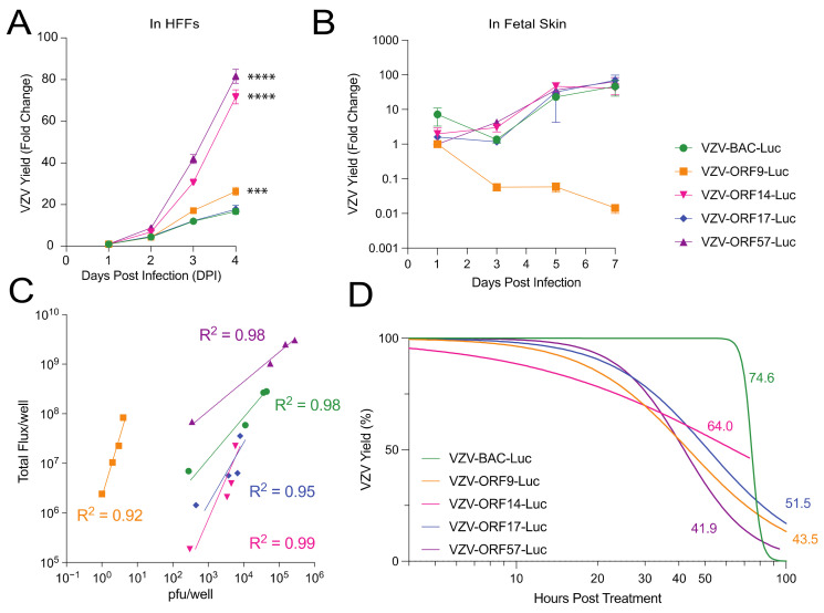
Figure 2.
Evaluation of VZV-BAC-Luc and VZV-ORFx-Luc growth kinetics and bioluminescence in tissue culture and human fetal skin. Cells and skin were infected with cell-free or cell-associated virus, respectively, and grown at 37 °C. Cell culture experiments (A,C,D) were performed in HFFs, while skin organ culture (B) was performed in fetal skin. (A,B) VZV yield was measured by bioluminescence imaging and calculated as the fold change from the average Total Flux (photons/sec/cm2/steradian), divided by the lowest Total Flux value (DPI 1 for cells, or DPI 1-3 for SOC). (C) Correlation coefficients of luciferase and virus plaque number were determined for each VZV reporter virus on the basis of the relationship of pfu/well to average Total Flux per well. (D) HFFs and VZV were co-cultured for approximately 40 h prior to foscarnet treatment (1 mM) to block viral DNA replication. Values next to each curve represent the time (in h) for bioluminescence to decrease by 50% after treatment started and are shown in the corresponding color for each virus (individual points omitted for clarity of graph). Each point represents the mean ± SEM. Statistical analyses included one-way ANOVA with Dunnett’s post hoc test (A,B, *** p < 0.001; **** p < 0.0001). Nonlinear regression analysis with (C) log–log line or (D) dose response—inhibition was used for best-fit lines. n = 3 biological replicates for cell-based assays; n = 6 biological replicates for skin organ culture.

