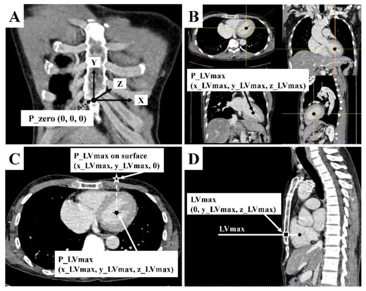
Figure 1.
Chest computed tomography measurement. (A) The zero point (P_zero: [0, 0, 0]) was defined as the midpoint of the xiphisternal junction. It is the palpable lower end of the sternum where all sternal body, xiphoid process, and costal margins meet. From P_zero, the leftward, upward, and into the thorax directions of the patient were designated as positive on x, y, and z axes, respectively, which formed right angles to one another. (B) The P_LVmax (x_LVmax, y_LVmax, z_LVmax) was the midpoint of maximal left ventricular diameter in coronal, sagittal, and axial view. Its 3D coordinate was identified by using the picture archiving and communicating system’s function. (C) Assuming that P_LVmax was located on the anterior chest surface (Z = 0) just vertically above that midpoint, defined as P_LVmax on the surface (x_LVmax, y_LVmax, 0). (D) The sagittal view of CCT was used to locate the maximal left ventricular diameter on the sternum, defined as LVmax (0, y_LVmax, z_LVmax).

