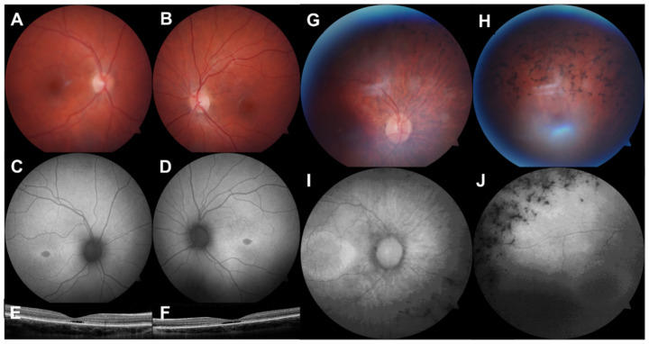
Figure 1.
Multimodal retinal imaging of 2 paediatric IRD cases. (A,B). Colour fundus photographs (FF450 plus, Carl Zeiss MediTec, Dublin, CA, USA) showing an altered foveal reflex. (C,D). Fundus autofluorescence images showing symmetrical hypoautofluorescence at the fovea with a hyperautofluorescent margin. (E,F). Optical coherence tomography (iVue 80, Optovue Inc., Fremont, CA, USA) showing outer retinal cavitation at the fovea. This 13-year-old female had Stargardt disease due to compound heterozygous pathogenic ABCA4 variants. (G,H). Colour fundus photographs of the right eye showing optic nerve head pallor, arteriolar attenuation, and intraretinal pigment migration. (I,J). Fundus autofluorescence showing a hyperautofluorescent macular ring and peripheral hypoautofluorescence. This 10-year-old female had autosomal dominant RP due to a likely pathogenic PRPF8 variant.

