Fig. 1.

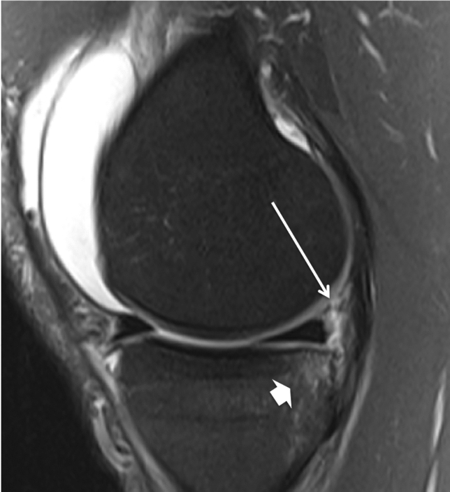
Sagittal fat-suppressed proton density weighted MRI shows a separation of the posteromedial capsule and the posterior horn of the medial meniscus (ramp lesion, long →) and bone oedema at the posterior medial tibial plateau (thick →)

Sagittal fat-suppressed proton density weighted MRI shows a separation of the posteromedial capsule and the posterior horn of the medial meniscus (ramp lesion, long →) and bone oedema at the posterior medial tibial plateau (thick →)