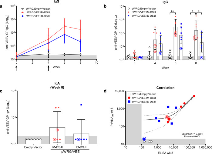
Fig. 1. pWRG/VEE delivered by IM- or ID-DSJI elicits anti-VEEV GP binding antibodies.
a Temporal anti-VEEV GP IgG responses and (b) total IgG antibody titers in vaccinated nonhuman primates (NHPs). c Total IgA antibody titers 4 weeks after the second vaccination in vaccinated NHPs. The shaded areas represent the assay limit based on the lowest sera dilution tested. Arrows represent vaccination time points. Data represent the group geometric mean ± geometric SD. *p < 0.05, **p < 0.01. d Correlation analysis of the VEEV ELISA and PsVNA80. For panels (a) and (c), p values were determined by one-way ANOVA with Tukey’s multiple comparison test. For panel (b), p values were determined by two-way ANOVA with Tukey’s multiple comparison. For panel (d), linear regression and 95% CI is shown as line and dotted lines, respectively. Circled symbols represent individual NHPs in VEEV-vaccinated groups that developed fever (defined as >3 SD above baseline lasting for more than 2 h) following VEEV aerosol challenge.

