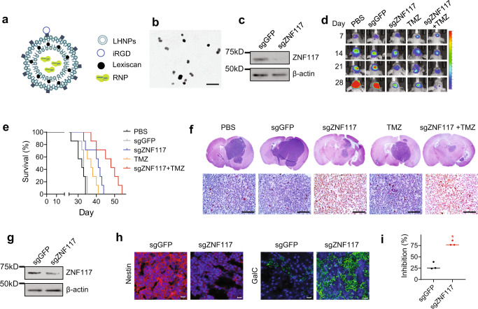
Fig. 6. Characterization of ZNF117 as a therapeutic target for GBM differentiation therapy.
a Schematics of LHNPs. b A representative image of LHNPs as captured by transmission electron microscopy (TEM). Experiments were performed in triplicate. Scale bar: 500 nm. c WB analysis of ZNF117 expression in PS30 cells treated with LHNPs loaded with Cas9 and sgGFP or sgZNF117. Experiments were performed in triplicate. d Representative images of tumors in the brain based on IVIS imaging. e Kaplan–Meier survival analysis of mice receiving the indicated treatments. f Representative H&E images of the brain isolated from mice receiving the indicated treatments. Experiments were performed in triplicate. Scale bar, 50 μm. g WB analysis of ZNF117 expression in residual tumors isolated from mice received the indicated treatments. Experiments were performed in triplicate. h Representative immunostaining images of residual tumors isolated from mice received the indicated treatments for expression of Nestin and GalC. Scale bar, 20 μm. i Inhibition of the proliferation of PS30 cells treated with Cas9 with the indicated sgRNA by TMZ (n = 3 biologically independent samples). Data are presented as mean ± SD. Black line represents mean, dots indicate values. Statistical differences were determined by two-tailed student’s t-test. *P-value < 0.05.

