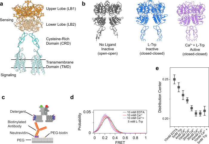
Fig. 1. CaSR is an intrinsically dynamic receptor, and ligands stabilize the active conformation.
a Ribbon representation the CaSR structure (PDB ID: 7DTW) colored to highlight the upper lobe (orange), lower lobe (pale orange), cysteine-rich domain (cyan), and the transmembrane domain (pale cyan). b Ribbon representation of the CaSR ectodomain in the Ioo, Icc, and Acc conformations (PDB IDs: 5K5T, 7DTU, and 7DTV respectively). c Schematic of single-molecule FRET experiments. d smFRET population histograms in the presence of 10 mM EDTA, 10 mM Ca2+, or 10 mM Ca2+ and 5 mM l-Trp. Data represent mean ± s.e.m. of n = 3 independent biological replicates. e Center of a single gaussian distribution fit to FRET histograms. Data represents the mean ± s.e.m. of n = 3 fits to independent biological replicates.

