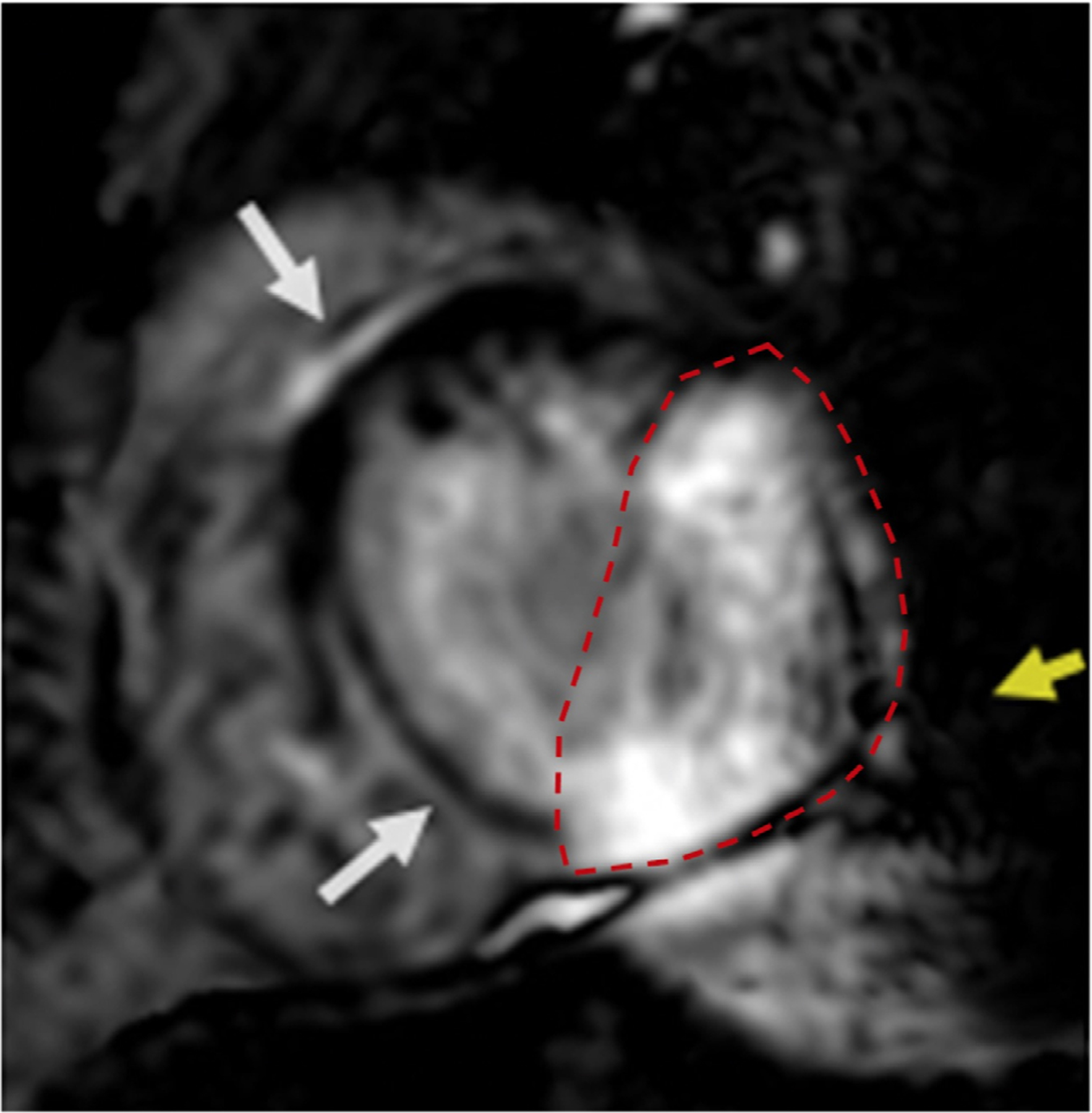
FIGURE 2:

Example of area of artifact estimation on an individual short-axis slice obtained using wideband late gadolinium enhancement (LGE) imaging in a patient with an S-ICD. (Dotted red line indicates the area of artifact; yellow arrow indicates mild signal void artifact; white arrows indicate mid septal LGE). ICD =implantable cardioverter-defibrillator; S-ICD = subcutaneous ICD.
