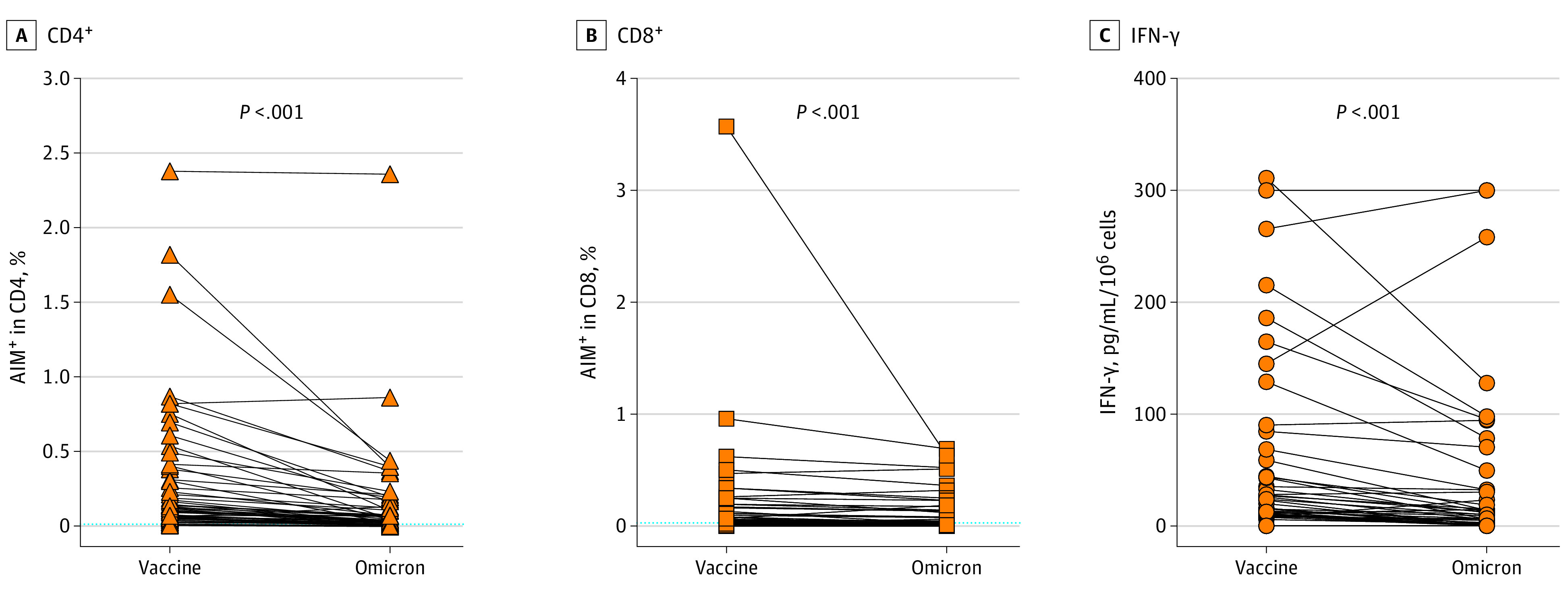
Figure 3. Reduced Recognition of Mutated Regions of the Spike Protein in the Omicron Variant.

Freshly isolated lymphocytes were incubated with peptide pools encompassing the mutated regions of the spike protein in the Omicron variant (Omicron), and with the reference peptide pool of the same region in the ancestral vaccine strain (Vaccine). Activated CD4+ (A) (CD69+ and CD40 ligand+) and CD8+(B) (CD69+ and CD137+) cells were identified by flow cytometry, and interferon (IFN)-γ production was measured in the supernatants (C). Background T-cell activation in paired unstimulated cultures was subtracted. Dotted lines indicate the threshold for positivity (median − 75th percentile of values from unstimulated cultures). Differences were assessed using Friedman rank sum test with Omicron exposure (vaccine / Omicron) as the random effect and participant ID (points) as the random effect.
