
Figure 1.
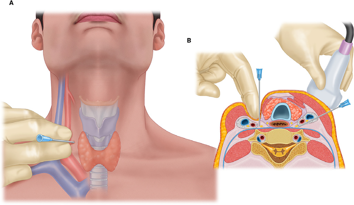
Panel A illustrates surface anatomy for stellate ganglion nerve block by the classic approach. The cricoid cartilage is palpated, and the vascular bundle is displaced laterally. The needle tip is inserted in a plane perpendicular to the insertion point on the skin. Panel B illustrates the cross section at the level of C6 showing the classic approach on the right side of the neck and ultrasound-guided approach on the left. Note the needle track is lateral to the vascular bundle and under the major vessels using inplane approach under ultrasound guidance. Proximity to various nerves, vessels, thyroid tissue, and esophagus can be appreciated in a cross-sectional view.