
Figure 2.
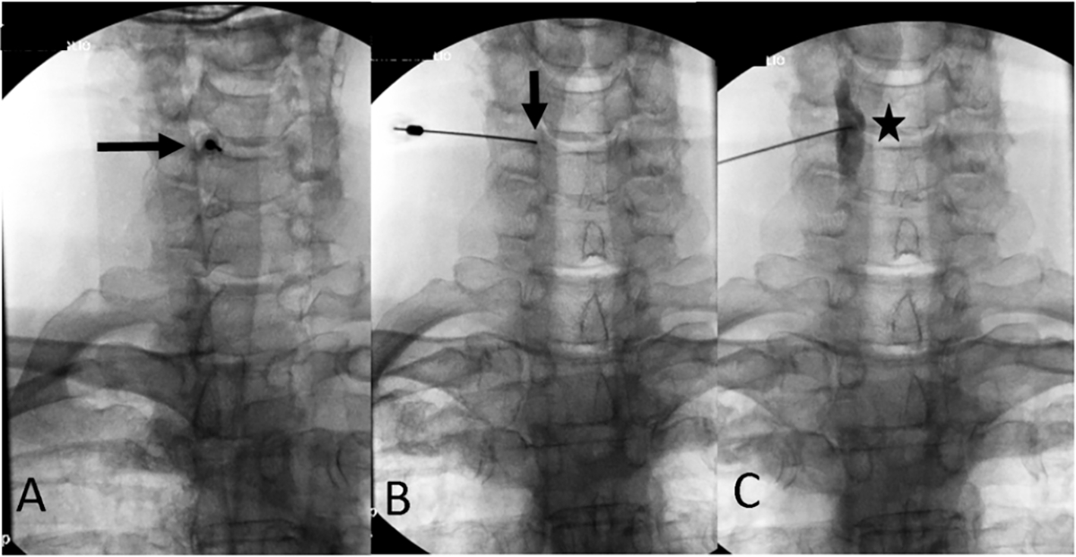
Fluoroscopy-guided oblique approach to stellate ganglion nerve block. Panel A illustrates the C5–C6 disc interspace in the right anterior oblique view. The needle is inserted coaxially to the beam targeting the C6 uncinate process (arrow). Panel B illustrates the final needle position (arrow) in the posteroanterior view. Panel C shows the contrast injection being performed to rule out vascular uptake. Contrast is seen to spread along the longus colli muscle in a craniocaudal fashion (star).