
Figure 3.
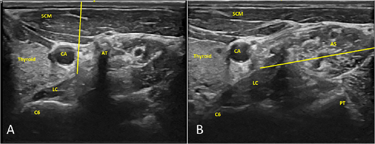
Ultrasound-guided imaging approach to stellate ganglion nerve block. Panel A illustrates the potential needle track (yellow line) with out-of-plane imaging. Panel B illustrates the potential needle track (yellow line) with inplane imaging. The ultrasound probe is placed on the right side of the neck at the C6 vertebral body level in the images. The yellow line is the trajectory for inplane needle ending at the prevertebral fascia covering the longus colli. AS, anterior scalene; AT, anterior tubercle; C6, cervical vertebra 6; CA, carotid artery; LC, longus colli muscle; PT, posterior tubercle; SCM, sternocleidomastoid muscle.