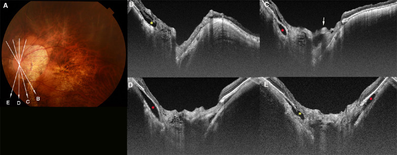
Figure 6.
Prelaminar schisis coexisting with ICC in an eye with PM. (A) A fundus photograph of the left eye of a 67-year-old woman with axial length of 29.9 mm. The arrows indicate the OCT scan lines in (B–E). (B–E) OCT images showing a meshwork-like prelaminar schisis in a wide area within the disc. A cross-section of an anteriorly protruding retinal vessel can be seen (arrow in C). Peripapillary retinoschisis and retinal detachment (yellow asterisks in B and E) can be seen in the superior region. Hypo-reflective area under the RPE (red asterisks in C–E), suggesting the presence of an ICC can also be seen.

