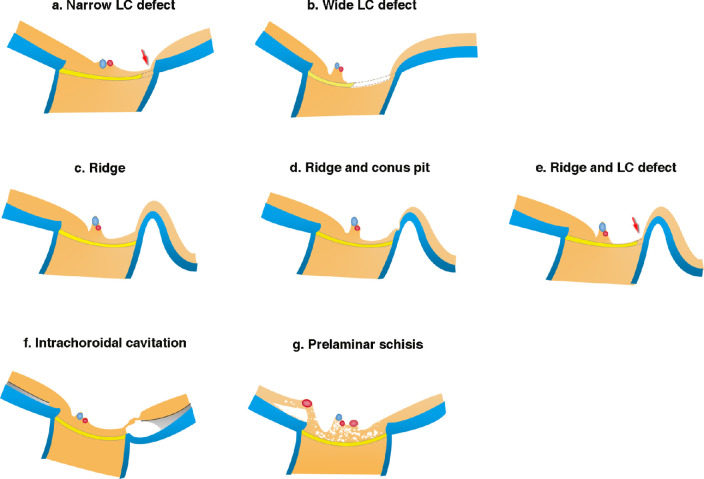
Figure 8.
Schematic drawing showing the different structural abnormalities in PM eyes. (a) Narrow LC defect (arrow) is observed as a pit or disinsertion of the LC. In some cases, the defect is deep and continues up to areas posterior to the LC. The overlying retina is thin, and in some cases almost absent. Surrounding retinal tissue gradually thins toward the site of LC defects. (b) A wide LC defect seems to extend up to one-half the LC width with obvious thinning of the prelaminar tissue. In eyes with very wide LC defects, the central trunk of the retinal vessels tends to shift toward the opposite side from the site of LC defects. (c) Ridge is an anterior protrusion of the sclera temporal to the optic disc. Retinal thinning is marked on the slope between disc margin and the ridge peak. (d) Ridge protrusion and conus pit commonly coexist, and the retinal tissue over the conus pit is extremely thin and almost absent. (e) Ridge and LC defect can coexist, and the retinal tissue is obviously thin on the slope of ridge and over the LC defect (arrow). (f) ICC with large area of fluid space. The overlying retinal tissue bridging the ICC can be extremely stretched and thinned. (g) Prelaminar schisis is mainly caused by the retinal vessels on the disc hanging anteriorly. Retinoschisis is also seen around the anteriorly protruded retinal vessels in the peripapillary area.

