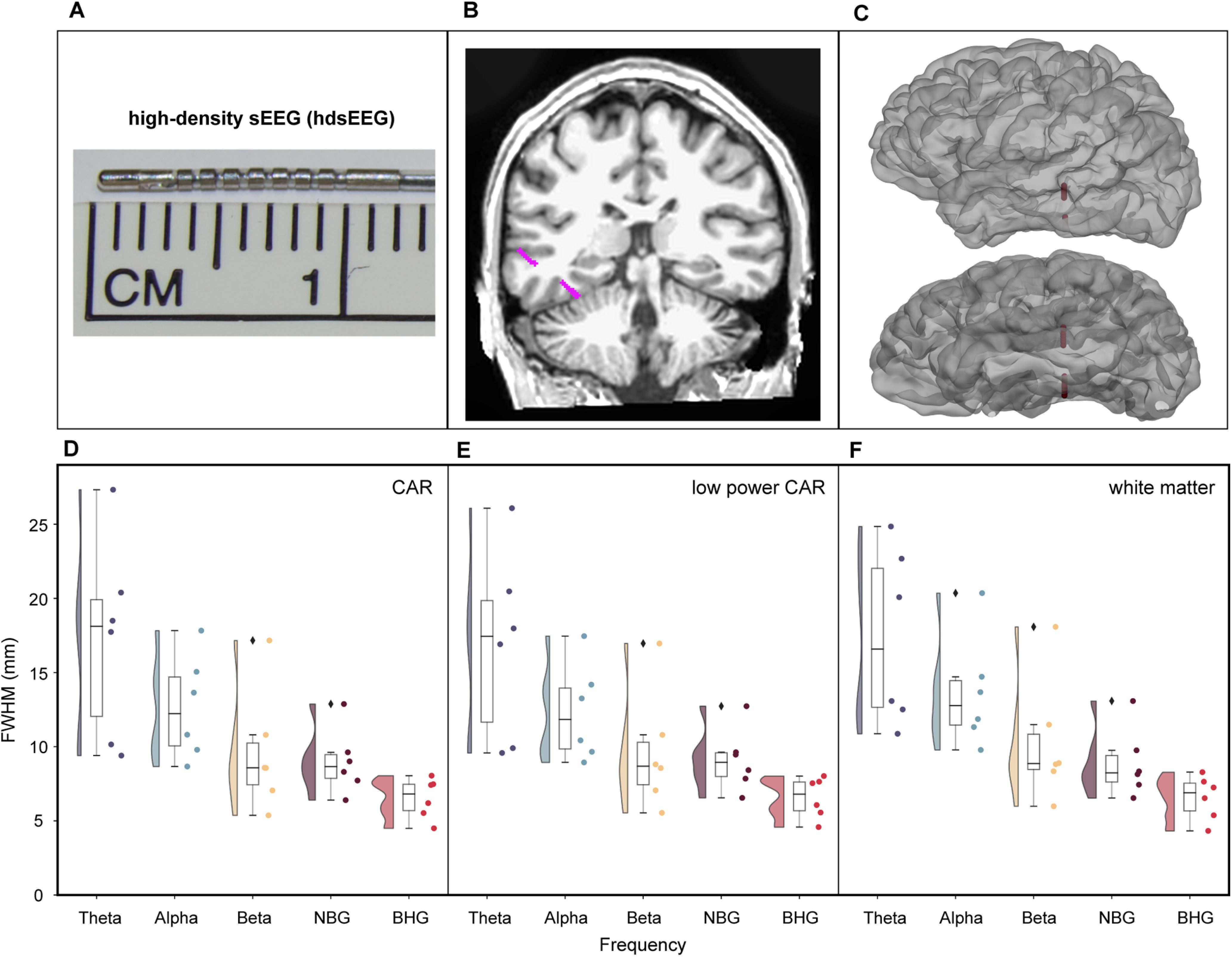
Figure 5.

Referencing scheme comparison across hdsEEG electrodes. hdsEEG electrodes are 0.5 mm in length and are on hybrid probes with sEEG 2 mm contacts (A). An exemplar hdSEEG electrode is shown in magenta in a coronal MRI slice (B) and in a surface reconstruction of the same patient (C). Average FWHM was calculated and plotted for each patient for hdsEEG electrode pairs in each frequency range of interest. For hdsEEG electrode pairs (6 patients; 153 electrodes; 1967 electrode pairs), average FWHM was compared using either CAR (D), low-power CAR (E) or white matter referencing schemes (F). Frequency ranges of interest: θ (4–8 Hz), α (8–13 Hz), β (13–30 Hz), NBG (30–60 Hz), BHG (70–150 Hz). NBG, narrowband γ; BHG, broadband high γ.
