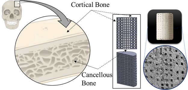
Figure 1.
Bimodal pore distribution bone scaffold. Interconnected porosity 3DP scaffolds are modeled after bone morphology and manufactured using powder bed binder jet printing. Bone contains cortical and trabecular bones (dense and spongy bones) which was the basis for the scaffold design used in this study. This bimodal pore distribution aids in increased mechanical properties by employing a dense exterior with a sustained interconnected porous interior that still allows for cell motility, nutrient flow, and vascularization. Dimensions of scaffolds are 7 mm diameter by 11 mm height for mechanical strength testing and 3 mm diameter by 5 mm height for in vivo implantation in a rat distal femur model.

