Figure 5.
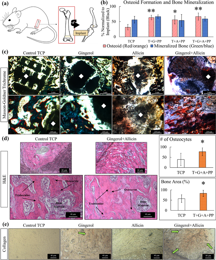
Week 4 in vivo analysis. (a) Schematic representation of in vivo rat distal femur model surgery. (b) Histomorphometry of osteoid formation and bone mineralization, normalized to the exposed implant area (black). Ginger extract (gingerol), garlic extract (allicin), and ginger + garlic extract (gingerol + allicin) show a significantly higher formation of osteoid compared to control TCP (n = 5, *p = 0.05, and **p = 0.01). (c) Optical microscopy images of quantified Masson-Goldner trichrome staining show prominent osteoid formation of ginger + garlic extract samples compared to the control. Color coding is as follows: implant (black marked with a white diamond), osteoid tissue (red/orange), and mineralized bone tissue (green/blue). Gaps in tissue growth are seen within control TCP scaffold implants at both timepoints comparatively to ginger + garlic extract-loaded TCP scaffolds. (d) H&E-stained tissues show healthy bone tissue, trabecular bone tissue, osteocytes, and bone marrow in all compositions. Ginger and garlic extracts both individually show support of osteogenesis, but results are more profound when they are used together. Ginger + garlic extract supports about a 90% increase in osteocytes and 30% increase in bone area (*, n = 5, p < 0.05). (e) Collagen staining shows an increase in collagen formation with ginger extract and even more so by ginger + garlic extract scaffolds in week 4. Collagen is indicated by the orange/yellow color and green arrow. Enhanced type I collagen can induce osteoblastic differentiation, thus aiding in osteogenesis and bone healing.

