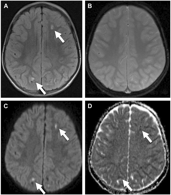
Fig. 1.

A 5-year-old boy with traumatic axonal injury at the level of the centrum semiovale. The lesions (arrows) are hyperintense on fluid-attenuated inversion recovery (FLAIR) (A), do not demonstrate appreciate susceptibility on T2*-weighted images (B), and show restricted diffusion on DWI (C) and apparent diffusion coefficient maps (D), reflecting acute injury.
