Figure 1.

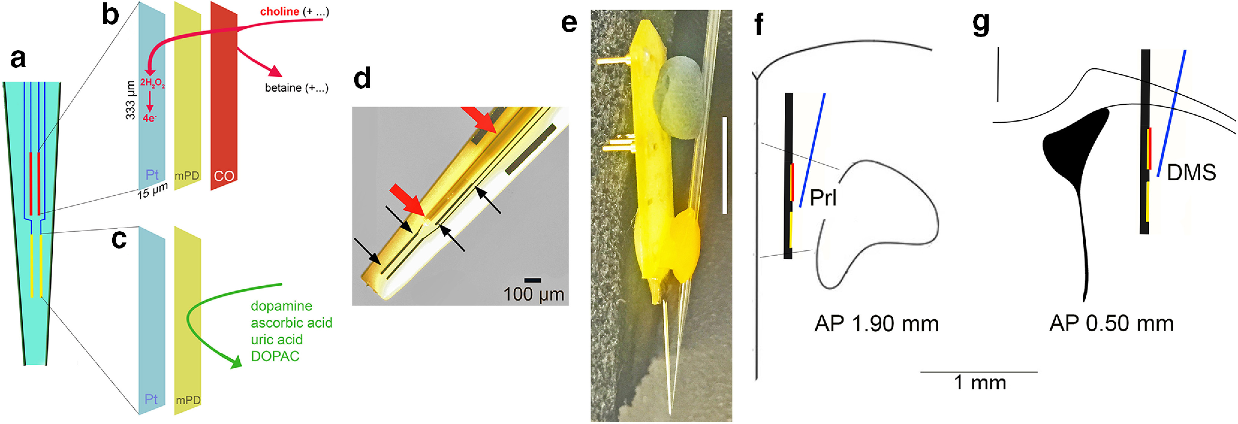
Diagram of the choline measurement scheme, the glass capillary attached to the electrode for pressure ejections, and placement of the assembly in the medial frontal cortex. a, Ceramic backbone with the four platinum-iridium (Pt/Ir) recording sites, each 333 µm long and 15 µm wide, organized in two pairs (see text for references). The upper pair was used to measure choline currents (red; see also b), while the lower pairs served as sentinels (yellow; see also c). The choline-sensing scheme is illustrated in abbreviated form in b, c. Choline is oxidized by choline oxidase (CO) that was immobilized onto the upper pair of recording sites. The resulting current, over background activity, was converted to choline concentration (μm). Before immobilization of CO, the nonconducting polymer m-(1,3)-phenylenediamine (mPD) was electroplated onto the recording sites to block the transfer of small electroactive organic molecules to the Pt site. Importantly, such mPD films do not block small species such as H2O2 from reaching the Pt surface (Huang et al., 1993; Mitchell, 2004). The lower pair of recording sites (c) were coated with mPD but not CO and thus served to record the concentration of electroactive interferents, which, despite the mPD barrier, still reached the Pt sites. A top view of the electrode in d shows the Pt recording sites (black arrows), with the upper left Pt site situated underneath a glass capillary (red arrows) used to pressure eject compounds into the recording area (100-µm scale inserted). The entire electrode/glass capillary assembly is shown from the side in e. Note that the Pt recording sites are not visible because of low resolution (1-cm scale vertically inserted on the right) but are located in the very tip of the ceramic backbone (see also the tip of the glass capillary). For the present experiments, the electrode/glass capillary assembly was inserted into the medial frontal cortex or the dorsomedial striatum of mice. f, g Coronal section of the frontal cortex (Prl, prelimbic cortex) and dorsomedial striatum, respectively, and the approximate placements and relative dimension of the assembly (AP, anterior-posterior coordinates).
