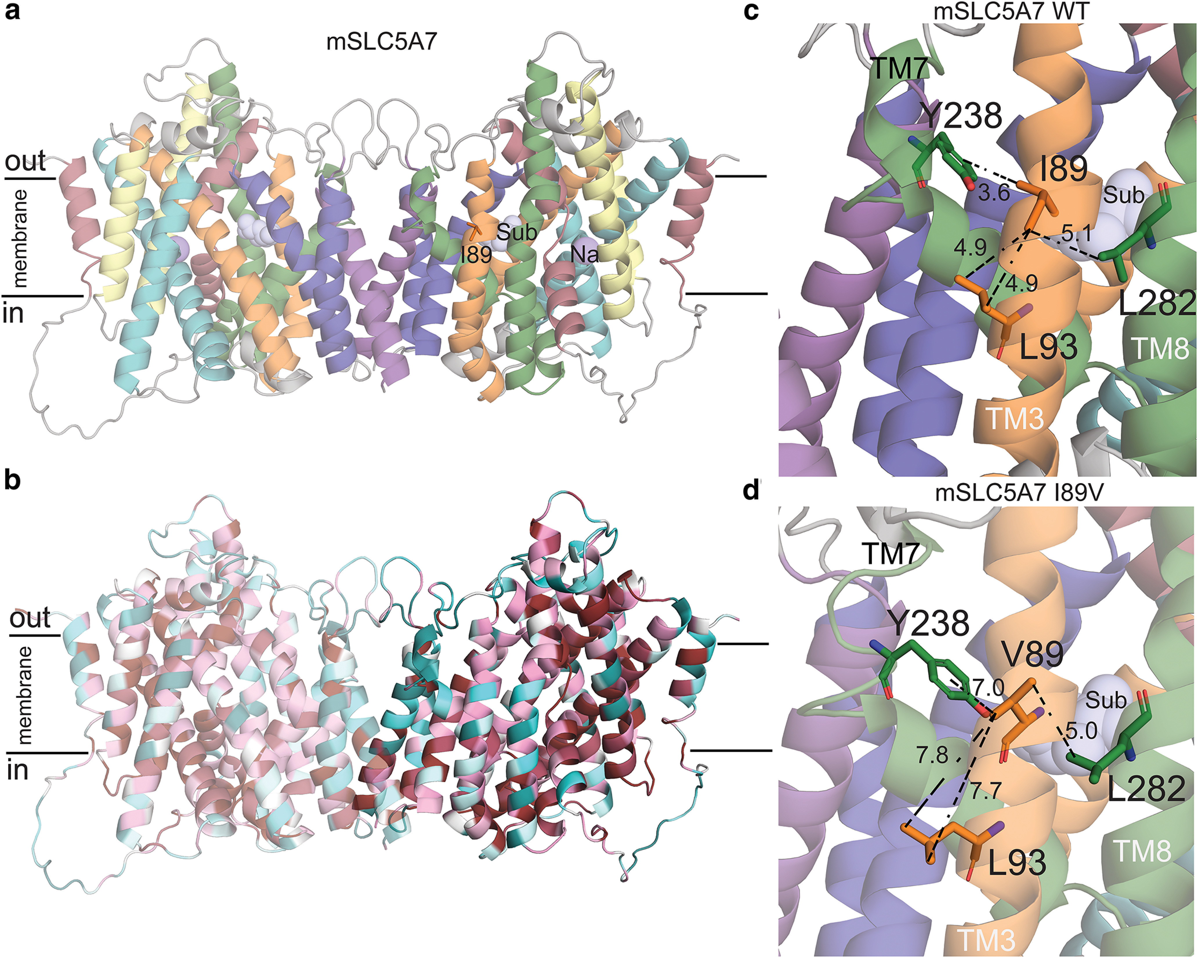
Figure 9.

Structural model of mouse CHT Val89. a, Structural model of mSLC5A7 in dimeric form colored according to the topology. I89, the sodium ion and substrate are highlighted as orange sticks and purple and light blue spheres, respectively. The position of the substrate and sodium ions where those obtained after structural superimposition of CHT model and vSGLT structure. b, Final model of mSLC5A7 where each residue is colored by its conservation score obtained with Consurf server (Ashkenazy et al., 2016). c, d, Close-up views of Ile89 and Val89 in their corresponding mSLC5A7 structures. Residues interacting with Ile89 are shown as sticks and the distances between sidechains are shown as dashed lines. The helices are colored as in a.
