
Extended Data Fig. 9. PARP1 activity is required to promote DNA repair and cell survival upon DNA damage.
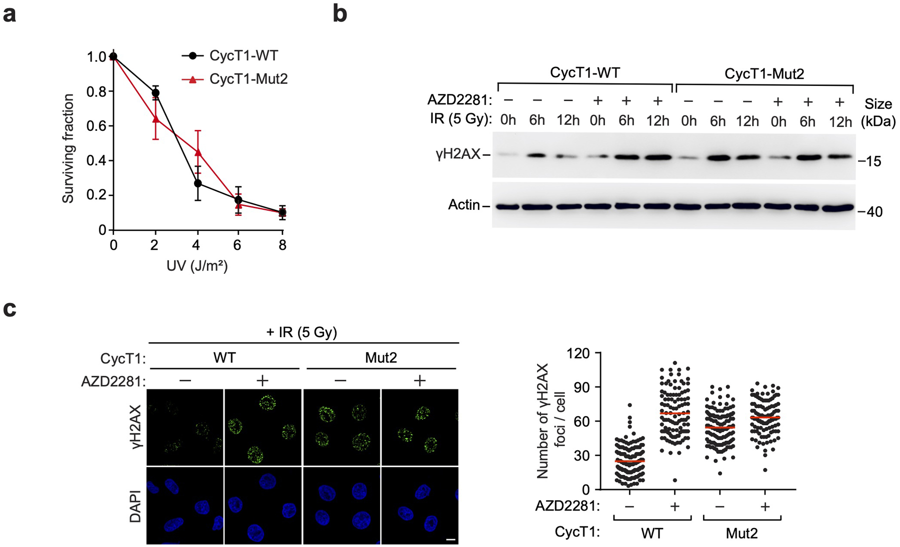
a, HeLa cells were depleted of CycT1 and reconstituted with WT or Mut2 CycT1. Cell viability was measured before or after the exposure to the indicated doses of UV. The error bars indicate mean ± s.d. with n = 3 biologically independent samples. b, Control or AZD2281-treated HeLa cells depleted of CycT1 and reconstituted with WT or Mut2 CycT1 were either mock treated or treated with IR (5Gy) and allowed to recover for the indicated time periods. WCE was examined by WB to detect γH2AX and actin. c, Control or AZD2281-treated HeLa cells depleted of CycT1 and reconstituted with WT or Mut2 CycT1 were analyzed by immunofluorescence staining at 12 hr after IR (5Gy) treatment with the anti-γH2AX antibody. DNA was counterstained by DAPI. Scale bar = 10 μM. Right: Quantification of the number of γH2AX foci per cell. Red lines indicate the mean γH2AX foci in each cell populations. n represents the number of cells examined in a representative assay out of 3 independent experiments: CycT1-WT-DMSO (n=148), CycT1-WT-AZD2281 (n=110), CycT1-Mut2-DMSO (n=138), CycT1-Mut2-AZD2281 (n=110). All Western blots are representative of three independent experiments. Gel source data are available online.