
Extended Data Fig. 2. CycT1 but not CDK9 in P-TEFb is PARylated in response to treatment with MNNG.
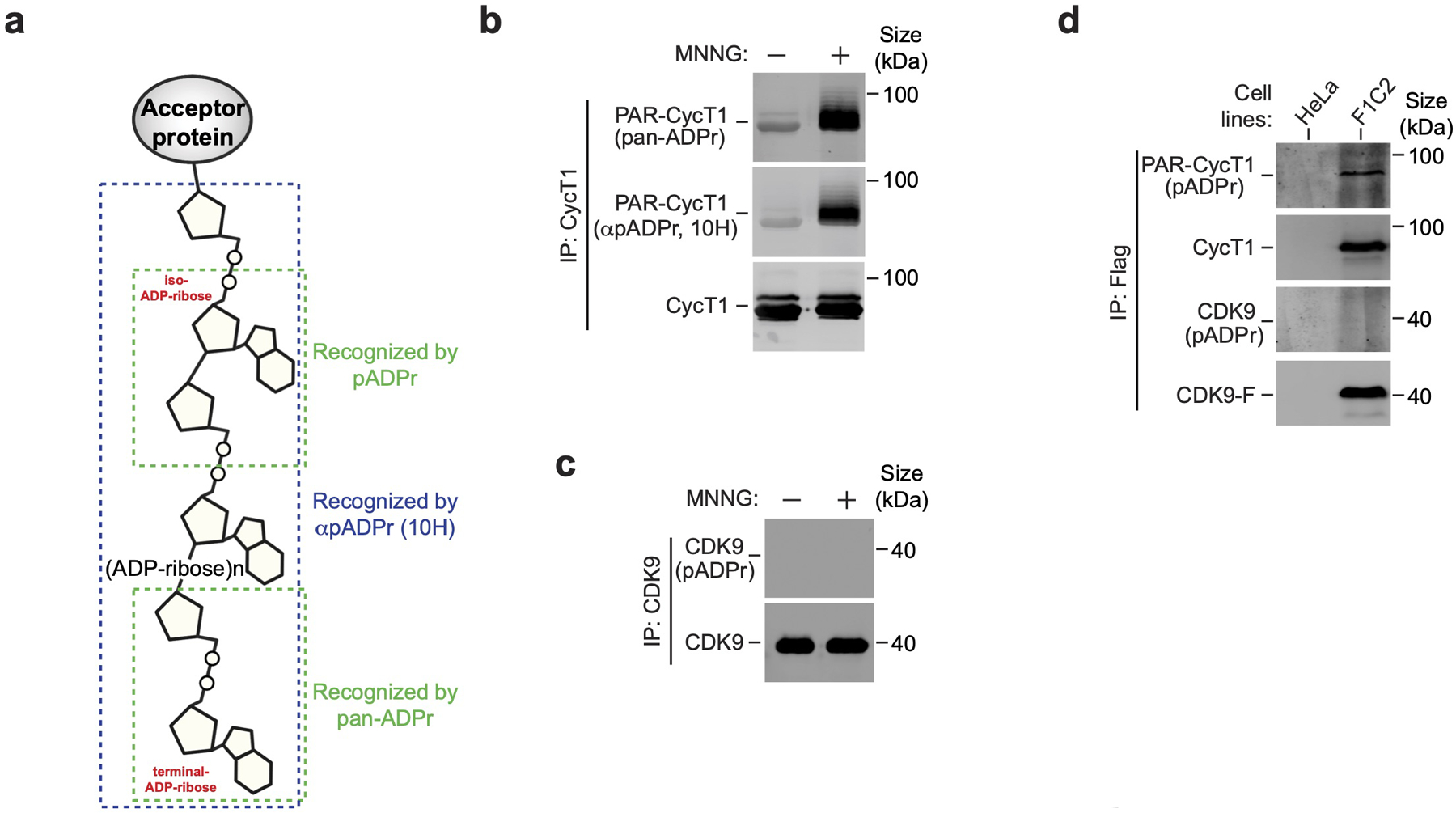
a, Schematic diagram depicting different regions on the PAR polymer that are recognized by pan-ADPr, αpADPr (10H) and pADPr, respectively. b, CycT1 was affinity-purified from HeLa cells either untreated or treated with MNNG and analyzed by Western blotting (WB) with pan-ADPr and αpADPr (10H). c, CDK9 was purified from HeLa cells, which were untreated or treated with MNNG, and analyzed by WB. d, HeLa or F1C2 cells stably expressing CDK9-F were treated with MNNG. Anti-Flag immunoprecipitates (IP) were analyzed by WB for the indicated proteins. All Western blots are representative of three independent experiments. Gel source data are available online.