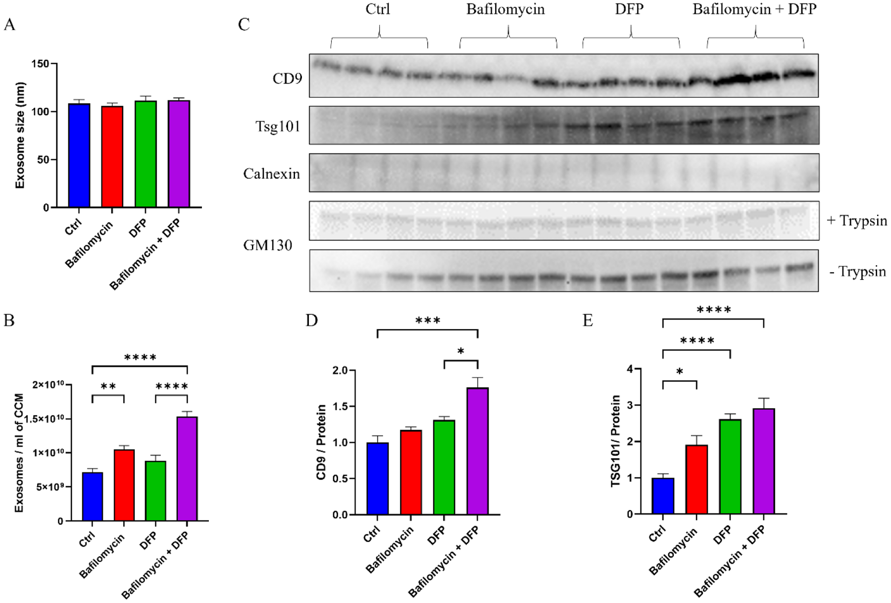
Figure 2. Effect of bafilomycin, DFP, or both on exosome secretion.

SH-SY5Y cells were treated with 50 nM bafilomycin for 24 hours, 1 mM DFP for 24 hours, or DFP for 24 hours with bafilomycin present for the final 16 hours. (A) Exosome size. (B) Number of exosome particles per ml of cell culture medium (CCM). (C) Western blots of exosome (CD9 and Tsg101), endoplasmic reticulum (calnexin), and Golgi body (GM130) markers. (D) CD9 protein levels. (E) TSG101 protein levels. Data represent means ± SEM, *p < 0.05, **p < 0.01, ***p < 0.001, ****p < 0.0001, as analyzed through one-way ANOVA with Tukey’s multiple comparisons test; significant changes relative to the no-treatment control (Ctrl), and between the DFP and bafilomycin+DFP conditions, are indicated.
