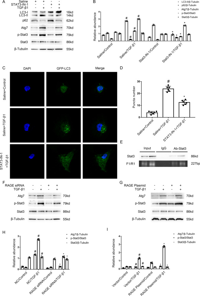
Fig. 4. The RAGE mediated the TGF-β1-induced autophagy in BUMPT cells via STAT3/Atg7 axis.
Immunoblotting results demonstrated that TGF-β1-induced the increasing of p-Stat3, Atg7, and LC3 II, and the reduction of p62 was notably attenuated by STAT3-IN-1, an STAT3 inhibitor. A Immunoblot analysis of LC3 & p62 & Atg7 & p-Stat3 & Stat3 in BUMPT cells. B Analysis of the grayscale image between them. #P < 0.05 versus Saline with the control group. *P < 0.05 versus Saline with the TGF-β1 group. C After GFP-LC3 transfection, HK-2 cells were transfected with or without STAT3-IN-1, followed by the treatment with TGF-β1 for 24 h. The punctate accumulation of GFP-LC3 was visualized under fluorescence microscopy. D The number of LC3 puncta per cell was calculated. #P < 0.05 versus Saline with the control group. *P < 0.05 versus Saline with the TGF-β1 group. E ChIP assays verified that a binding sites (a 227 bp fragment) of STAT3 existed in the promoter region of Atg7. The plasmid and siRNA of RAGE were transfected into BUMPT cells and treated with or without 5 ng/ml TGF-β1 for 24 h. F, G Immunoblot analysis of Atg7 & p-Stat3 & Stat3 in BUMPT cells. H, I Analysis of the grayscale image between them. #P < 0.05 versus NC or Vector with the control group. *P < 0.05 versus Saline with the TGF-β1 group. Each experiment A, C, F, G was repeated six times independently with similar results. B, H, I indicate the statistical Student’s t test used (means ± s.d., n = 6, P < 0.05).

