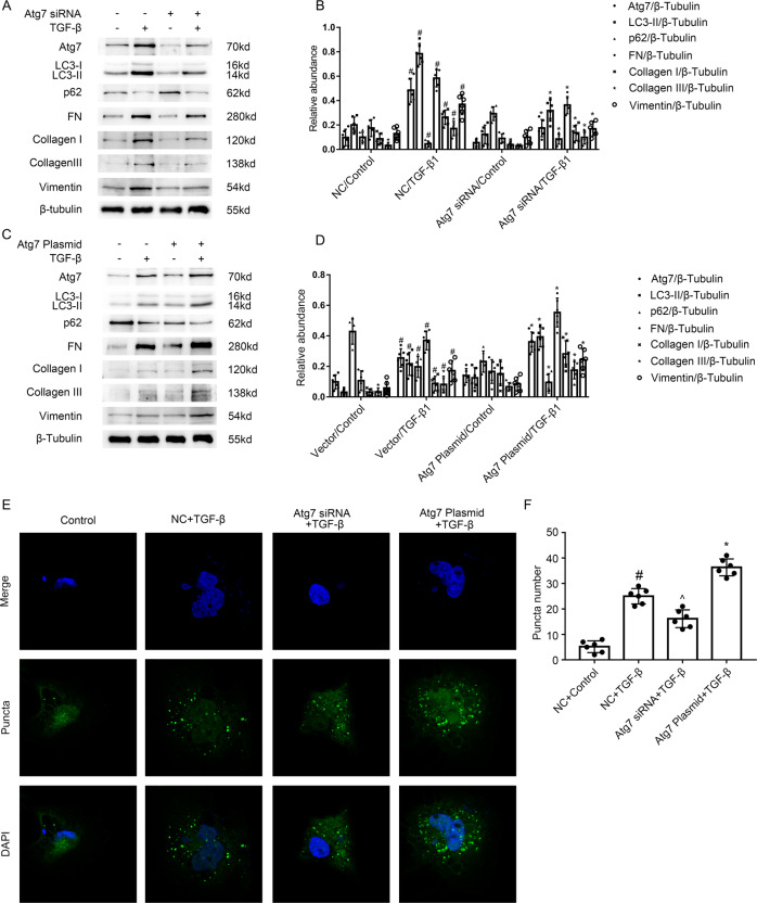
Fig. 5. Atg7 mediated the TGF-β1-induced autophagy in BUMPT cells.
The plasmid and siRNA of Atg7 were transfected into BUMPT cells and treated with or without 5 ng/ml TGF-β1 for 24 h. A, C Immunoblot analysis of Atg7 & LC3 & p62 & FN & Collagen I & Collagen III and Vimentin in BUMPT cells. B, D Analysis of the grayscale image between them. #P < 0.05 versus NC or Vector with the control group. *P < 0.05 versus NC or Vector with the TGF-β group. E After GFP-LC3 transfection, HK-2 cells were transfected with or without the plasmid or siRNA of ATG7, followed by the treatment with TGF-β1 for 24 h. The punctate accumulation of GFP-LC3 was visualized under fluorescence microscopy. F The number of LC3 puncta per cell was calculated. #P < 0.05 versus NC or Vector with the control group. ^P < 0.05 versus NC or Vector with the TGF-β group. *P < 0.05 versus NC or Vector with the TGF-β group. Each experiment A, C, E was repeated six times independently with similar results. B, D, F indicate the statistical Student’s t test used (means ± s.d., n = 6, P < 0.05).

