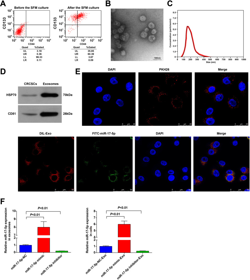
Fig. 1. CRC stem cell sorting and exosome identification.
A CD133+CD44+ CRCSC were sorted by flow cytometry; B the exosome morphology was observed using a TEM; C the diameter distribution and concentration of exosomes were detected by NTA; D the exosome markers HSP70 and CD81 in CRCSCs and exosomes were detected by western blot analysis; E the entry of exosomes that labeled with fluorescent PKH67 to HCT116 cells was observed by immunofluorescence microscopy; the co-localization of labeled fluorescent FITC-miR-17-5p and exosomes in HTC116 cells was observed by immunofluorescence microscopy; FITC-miR-17-5p-labeled exosomes were green; DAPI-stained nuclei were blue; DIL-labeled exosomes were red; F miR-17-5p expression in cells and exosomes detected using RT-qPCR. The cell experiments were repeated three times; P < 0.05; the measurement data were expressed as mean ± standard deviation.

