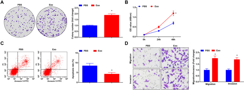
Fig. 2. CRCSC-exos promote malignancy of HCT116 cells.
A Proliferation of HCT116 cells detected using colony formation assay after exosome treatment; B proliferation of HCT116 cells detected using CCK-8 assay after exosome treatment; C apoptosis of HCT116 cells detected using flow cytometry after exosome treatment; D migration and invasion of HCT116 cells detected using Transwell assay (magnification: ×200) after exosome treatment. The cell experiments were repeated three times; *P < 0.05 vs the PBS group; the measurement data were expressed as mean ± standard deviation.

