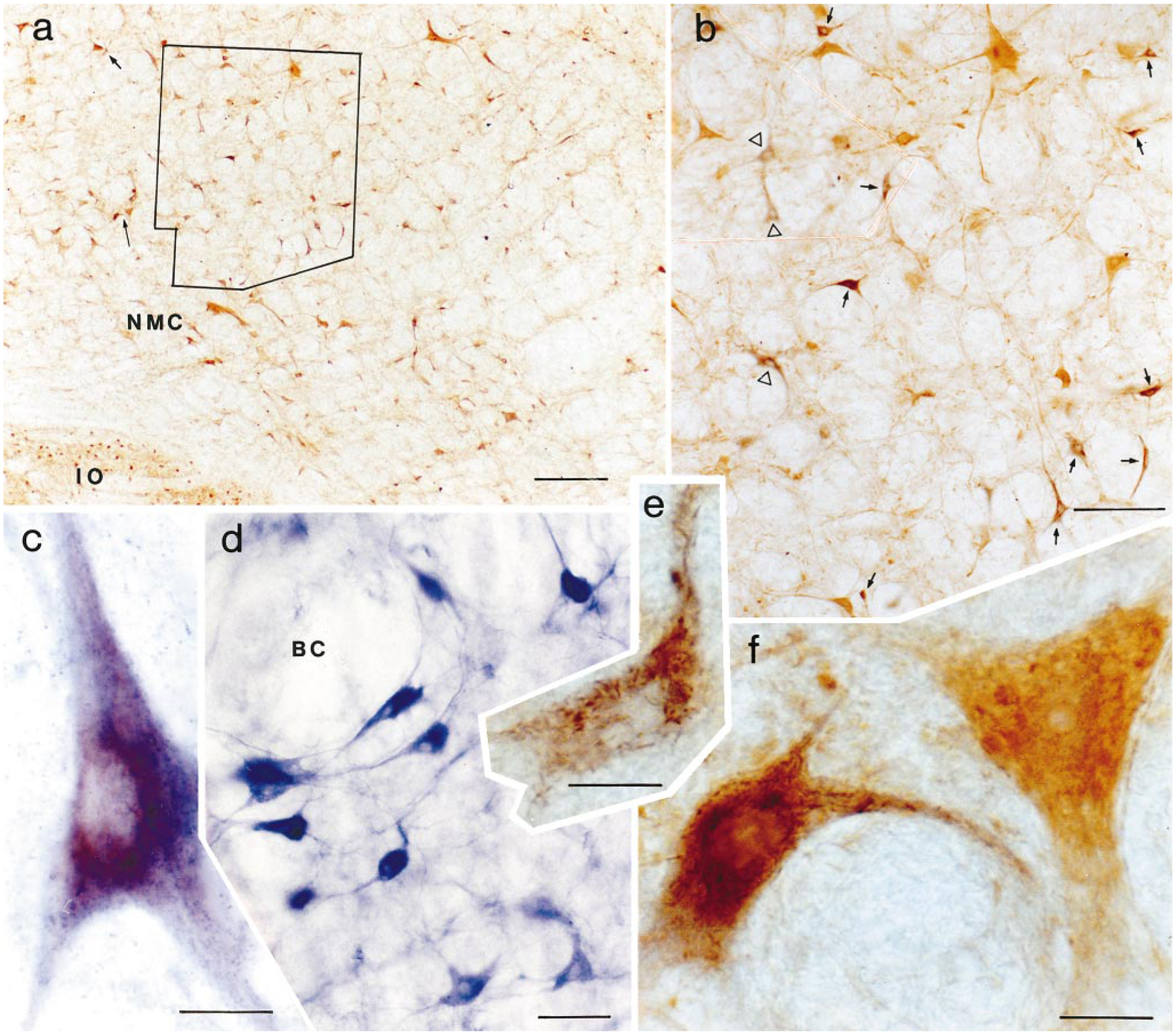
Fig. 3.

Photomicrographs showing wheat germ agglutinin–horseradish peroxidase (WGA-HRP) cells and cells double labeled with either glutamate or nicotinamide adenine dinucleotide phosphate-diaphorase (NADPH-d). a: Low magnification photomicrograph taken from the nucleus magnocellularis contralateral to the injection site. The section level is also shown in Figure 5, part 2 as level P10.0. b: Higher magnification photomicrograph taken of the area outlined within a. Many neurons labeled with WGA-HRP (open triangles) or WGA-HRP and glutamate (arrows) could be seen in this area. c: Double-labeled NADPH-d and WGA-HRP neurons in the nucleus paragigantocellularis contralateral to the injection site. Crystalline tetramethyl benzidine products are superimposed on the light-blue NADPH-d reaction. d: NADPH-d-positive neurons in the pedunculopontine nucleus. NADPH-d-positive neurons in the nucleus are large and deep blue and have several processes. e: High magnification photomicrograph shows WGA-HRP-labeled neuron shown in a (short arrow). f: High magnification photomicrograph showing homogenous yellow glutamate-immunoreactive neuron and double-labeled glutamate/WGA-HRP neuron shown in a (long arrow). For abbreviations, see list. Scale bars = 200 μ in a, 100 μ in b, 20 μ in c–f.
