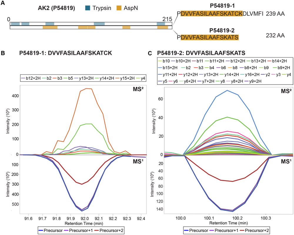
Figure 5. Isoform identification.
(A) Schematic of sequence coverage of AK2. Isoform 2 differs from isoform 1 by the replacement of a seven amino acid sequence at the C-terminal with serine. Regions of AK2 detected in the multiplexed mixture with trypsin (blue) or AspN (yellow) are highlighted. (B) XIC of isoform 1 C-terminal peptide. (C) XIC of isoform 2 C-terminal peptide. XICs for these peptides across all replicates are included as Supplemental Figs. 3A and B.

