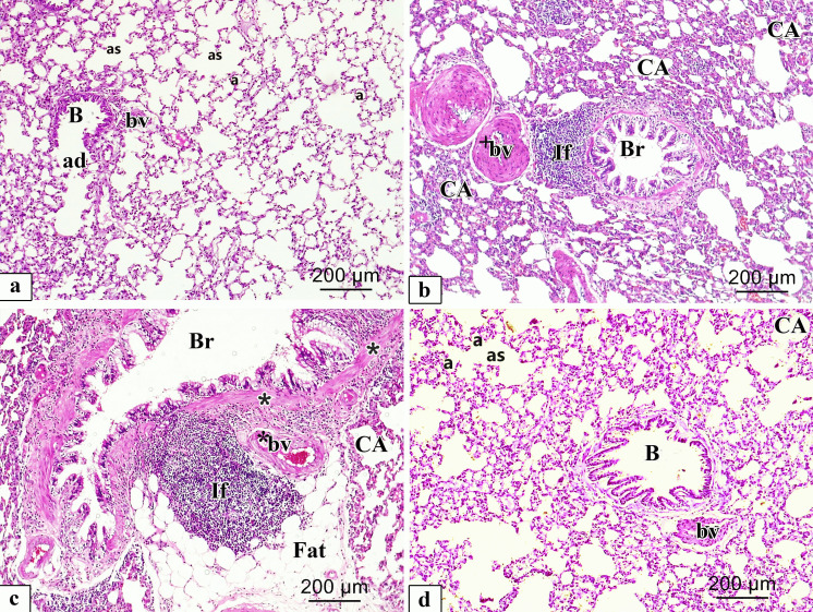
Fig. 6.
Representative microscopic images of H&E-stained sections of rat lung from different experimental groups. a. Control; b, c. CP and d. ALO + CP groups. large bronchiole (B), alveolar duct (ad), alveolar sac (as), alveoli (a), blood vessel (bv), collapsed alveoli (CA), Thick wall (*) bronchus (Br), extensive peri bronchial cellular infiltration (If), vascular congestion (*bv), thick wall blood vessel (+ bv), fatty cellular infiltrations (Fat)

