
Figure 5.
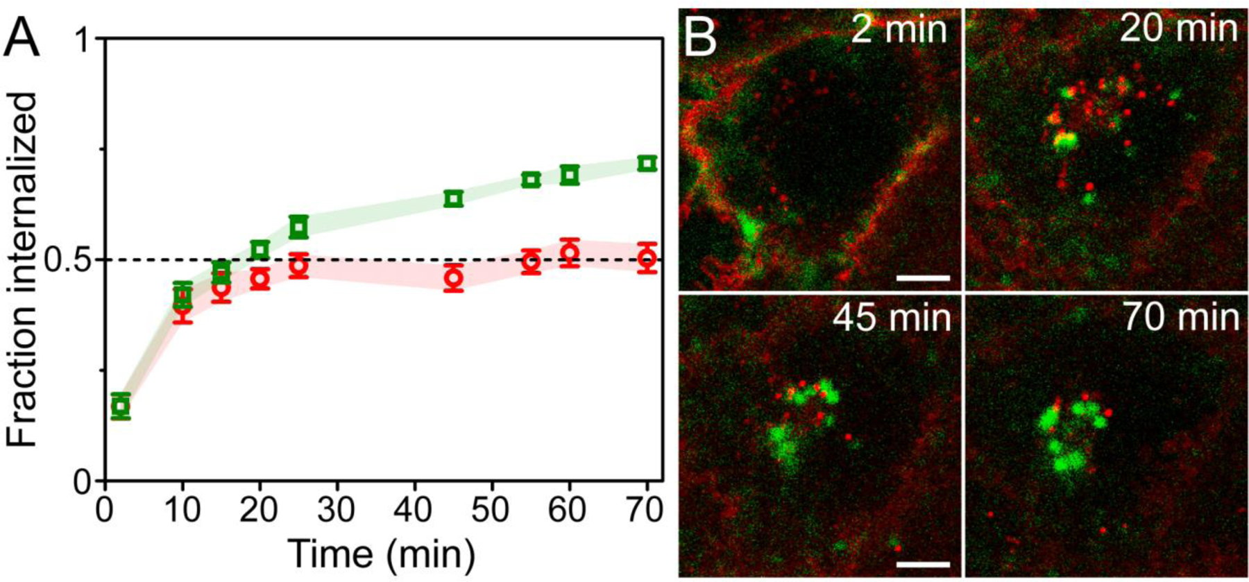
Differences of internalization and recycling between anti-uPAR scFv-UCNPs and a lipophilic membrane stain in breast cancer cells. (A) Fraction internalized of 2G10 scFv-UCNP immunoconjugates in live MDA-MB-231 cells during incubation at 37°C (mean ± SEM; n = 6 cells). (B) Confocal images of anti-uPAR-UCNPs (green squares) and lipophilic membrane fluorophore (red circles) in live MDA-MB-231 cells showing differences in intracellular localization between probes. Details from frames at 2, 20, 45 and 70 min of full 70-min time-lapse (Supplementary movie 1) are shown. Scale bars are 10 μm.